Ficheiro:Amebiasis LifeCycle.gif

Amebiasis_LifeCycle.gif (435 × 548 píxeis, tamanho: 27 kB, tipo MIME: image/gif)

Esta imagem de um diagrama (ou todas as imagens neste artigo ou categoria) deveriam ser recriadas usando gráficos vectoriais, como ficheiros SVG. Isto tem várias vantagens; veja as Commons:Media for cleanup|imagens para rever para mais informações. Se já criou um ficheiro SVG desta imagem, por favor, carregue-o. Depois do novo ficheiro SVG ter sido carregado, substitua aqui esta predefinição pela predefinição {{vector version available|nome da nova imagem.svg}}.

Descrição do ficheiro

Descrição
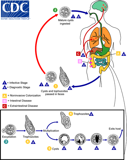
English: Cysts and trophozoites are passed in feces The Number 1. Cysts are typically found in formed stool, whereas trophozoites are typically found in diarrheal stool. Infection by Entamoeba histolytica occurs by ingestion of mature cysts The Number 2 in fecally contaminated food, water, or hands. Excystation The Number 3 occurs in the small intestine and trophozoites The Number 4 are released, which migrate to the large intestine. The trophozoites multiply by binary fission and produce cysts The Number 5, and both stages are passed in the feces The Number 1. Because of the protection conferred by their walls, the cysts can survive days to weeks in the external environment and are responsible for transmission. Trophozoites passed in the stool are rapidly destroyed once outside the body, and if ingested would not survive exposure to the gastric environment. In many cases, the trophozoites remain confined to the intestinal lumen (The Letter A: noninvasive infection) of individuals who are asymptomatic carriers, passing cysts in their stool. In some patients the trophozoites invade the intestinal mucosa (The Letter B: intestinal disease), or, through the bloodstream, extraintestinal sites such as the liver, brain, and lungs (The Letter C: extraintestinal disease), with resultant pathologic manifestations. It has been established that the invasive and noninvasive forms represent two separate species, respectively E. histolytica and E. dispar. These two species are morphologically indistinguishable unless E. histolytica is observed with ingested red blood cells (erythrophagocystosis). Transmission can also occur through exposure to fecal matter during sexual contact (in which case not only cysts, but also trophozoites could prove infective).
Data Data desconhecida
Origem https://www.cdc.gov/dpdx/amebiasis/index.html
Autor CDC
Outras versões

Licenciamento

Public domain
Esta imagem é um trabalho dos Centers for Disease Control and Prevention, parte do Departamento de Saúde e Serviços Humanos dos Estados Unidos da América, tirada ou feita durante o curso de uma tarefa oficial de um funcionário. Como trabalho do Governo Federal dos Estados Unidos da América, a imagem está no domínio público.

Legendas

Adicione uma explicação de uma linha do que este ficheiro representa

Elementos retratados neste ficheiro

retrata

6b2e40a81cf8320224551c870aca4af1ab73029f

27 701 byte

548 pixel

435 pixel

Histórico do ficheiro

Clique uma data e hora para ver o ficheiro tal como ele se encontrava nessa altura.

Data e horaMiniaturaDimensõesUtilizadorComentário
atual21h26min de 21 de julho de 2015Miniatura da versão das 21h26min de 21 de julho de 2015435 × 548 (27 kB)CFCFUser created page with UploadWizard

A seguinte página usa este ficheiro:

Utilização global do ficheiro

As seguintes wikis usam este ficheiro: