Ficheiro:Attic Cholesteatoma.jpg
Dimensões desta antevisão: 605 × 600 píxeis. Outras resoluções: 242 × 240 píxeis | 484 × 480 píxeis | 775 × 768 píxeis | 1 033 × 1 024 píxeis | 2 299 × 2 279 píxeis.
Imagem numa resolução maior (2 299 × 2 279 píxeis, tamanho: 231 kB, tipo MIME: image/jpeg)
|
|
Esta imagem provém do Wikimedia Commons, um acervo de conteúdo livre da Wikimedia Foundation que pode ser utilizado por outros projetos.
|
Descrição do ficheiro
| Descrição |
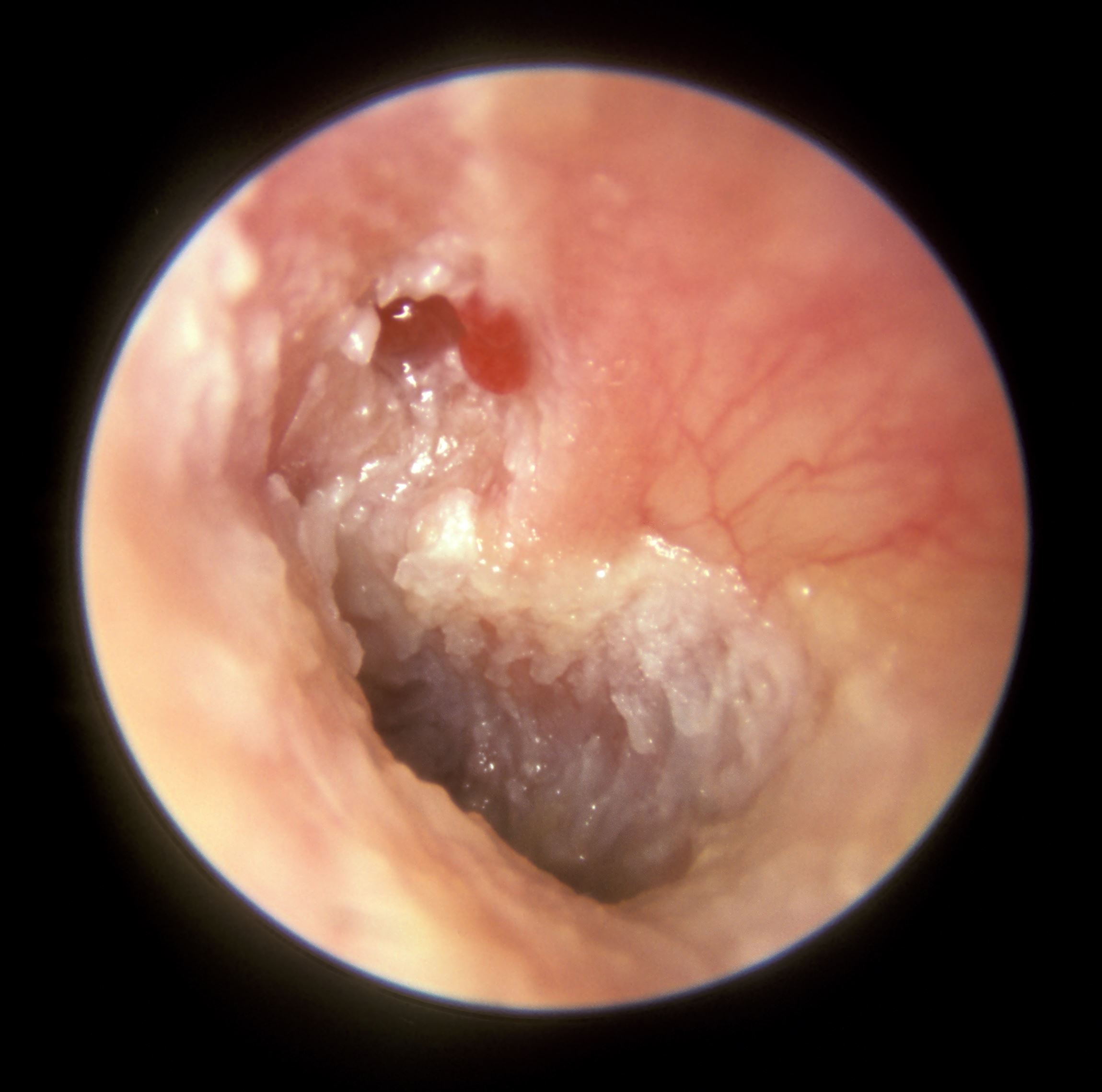
English: The cherry red small mass of granulation tissue arising from the Pars Flaccida of this left tympanic membrane strongly suggests that there is an underlying cholesteatoma in the attic area.
Deep to the granulation tissue there was a perforation through the pars flaccida which connected with the underlying attic cholesteatoma. Note that continuing discharge from the granulation tissue has caused the surface epithelia of the tympanic membrane to become edematous and thickened giving the surface a thickened whitish appearance. |
| Data | |
| Origem | Obra do próprio |
| Autor | Michael Hawke MD |
Licenciamento
Eu, titular dos direitos de autor desta obra, publico-a com a seguinte licença:
A utilização deste ficheiro é regulada nos termos da licença Creative Commons Atribuição 4.0 Internacional.
- Pode:
- partilhar – copiar, distribuir e transmitir a obra
- recombinar – criar obras derivadas
- De acordo com as seguintes condições:
- atribuição – Tem de fazer a devida atribuição da autoria, fornecer uma hiperligação para a licença e indicar se foram feitas alterações. Pode fazê-lo de qualquer forma razoável, mas não de forma a sugerir que o licenciador o apoia ou subscreve o seu uso da obra.
Histórico do ficheiro
Clique uma data e hora para ver o ficheiro tal como ele se encontrava nessa altura.
| Data e hora | Miniatura | Dimensões | Utilizador | Comentário | |
|---|---|---|---|---|---|
| atual | 13h54min de 7 de junho de 2015 | | 2 299 × 2 279 (231 kB) | DocHawke | User created page with UploadWizard |
Utilização local do ficheiro
A seguinte página usa este ficheiro: