Ficheiro:FluorescentCells.jpg

FluorescentCells.jpg (512 × 512 píxeis, tamanho: 56 kB, tipo MIME: image/jpeg)

Descrição do ficheiro

Descrição
English: a

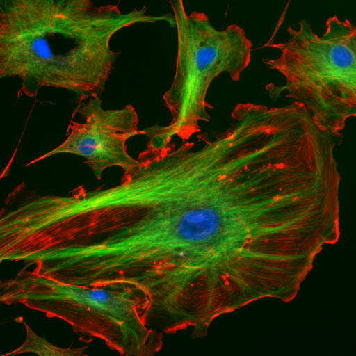
This image is made from a Molecular Probes demo slide:

  • Cells: bovine pulmonary arthery endothelial cells
  • Blue: nucleus stained with DAPI
  • Green: Tubulin (microtubles) stained with antibody Bodipy FL goat anti-mouse IgG (Indirect fluorescent antibody stain)
  • Red: F-Actin stained with Texas Red X-Phalloidin
Deutsch: Endothelzellen aus der Inneren Wand (Endothel) von Lungenarterien des Rindes unter dem Mikroskop. Die Zellkerne sind mit DAPI blau markiert. Die Mikrotubuli wurden über einen Antikörper grün markiert. Mit rot fluoreszierendem Phalloidin wurden die Aktinfilamente markiert.
Français : Cellulles endothéliales vues au microscope. En bleu, noyaux marqués au DAPI. En vert, microtubules marqués par un anticorps. En rouge, actine marquée à la phalloïdine.
Magyar: Fluoreszcenciamikroszkópos felvétel marha tüdőartéria endotélsejtjeiről (Molecular Probes FluoCells prepared slide #2 (F14781)). A sejtmagok DAPI-val vannak festve (kék), a mikrotubulusokhoz anti-α-tubulin egéranitest, ahhoz pedig BODIPY FL-el jelölt anti-egér kecske-IgG van kapcsolva (zöld), míg az aktin filamentumok Texas Red-X-el kapcsolt falloidinnal vannak jelölve (vörös). A kép három felvétel szuperpozíciójával készült. Hamis színek.
Lietuvių: Citoskeletas. Aktino filamentai – raudona, mikrovamzdeliai – žalia, branduolys – mėlyna spalva.
Română: Sub microscop Celule endoteliale . microtubulii sunt de culoare verde, iar filamentele de actină sunt roşii, pe când nucleul celulei este colorat albastru
Русский: Цитоскелет эукариот. Актиновые микрофиламенты окрашены в красный (фаллоидином, связанным с TRITC), микротрубочки — в зеленый (антителами, связанными с FITC), ядра клеток — в голубой цвет (DAPI). Клетки эндотелия лёгочной артерии быка.
Українська: Цитоскелет еукаріот. Актинові мікрофіламенти забарвлені в червоний колір, мікротрубочки — в зелений, ядра кліток — в блакитний
Origem http://rsb.info.nih.gov/ij/images/
Autor
Permissão
(Reutilizar este ficheiro)
example image from the ImageJ-Programmpaket (public domain)

Original file

This image has been taken from the German Wikipedia

The original uploader is de:Benutzer:Jan R. The original upload was at 4th December 2005.

Original description

This image is made from a Molecular Probes demo slide:

Cells: bovine pulmonary arthery endothelial cells
Blue: nucleus stained with DAPI
Green: Tubulin stained with Bodipy FL goat anti-mouse IgG
Red: F-Actin stained with Texas Red X-Phalloidin

(description from [1])

Quelle: Beispielsbild aus dem ImageJ-Programmpaket (public domain), siehe http://rsb.info.nih.gov/ij/

Licenciamento

Public domain
Esta obra encontra-se no domínio público nos Estados Unidos por ser uma obra do Governo Federal dos EUA nos termos do Título 17, Capítulo 1, Secção 105 do US Code. Veja Copyright. Nota: Este estatuto apenas se aplica a obras do Governo Federal, e não a obras de nenhum estado, condado, subdivisão ou município.
Este ficheiro foi considerado livre de restrições conhecidas devidas a direitos de autor, incluindo todos os direitos conexos.

Legendas

Adicione uma explicação de uma linha do que este ficheiro representa

Elementos retratados neste ficheiro

retrata

image/jpeg

Histórico do ficheiro

Clique uma data e hora para ver o ficheiro tal como ele se encontrava nessa altura.

Data e horaMiniaturaDimensõesUtilizadorComentário
atual15h07min de 24 de março de 2006Miniatura da versão das 15h07min de 24 de março de 2006512 × 512 (56 kB)Splette{{Information |Description = Endothelial cells under the microscope. Nuclei are stained blue with DAPI, microtubles are marked green by an antibody and actin filaments are labelled red with phalloidin. |Source = http://rsb.info.nih.gov/ij |Date = |Author

As seguintes 2 páginas usam este ficheiro:

Utilização global do ficheiro

As seguintes wikis usam este ficheiro:

Ver mais utilizações globais deste ficheiro.
Metadados